Dr. Maron's dental office
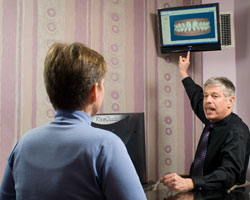
![]() Dr Maron in the consult room explaining the Invisalign treatment plan to the patient
Dr Maron in the consult room explaining the Invisalign treatment plan to the patient
- Invisalign computer-generated orthodontic treatment plans—Invisalign® is a system of moving teeth in small increments with clear plastic aligners. The Invisalign treatment plan plots the step-by-step movement of the teeth in a series of computer-generated images. This plan enables us to show you visually what the proposed orthodontic treatment should accomplish. With conventional orthodontics, you cannot see what the final result will be.
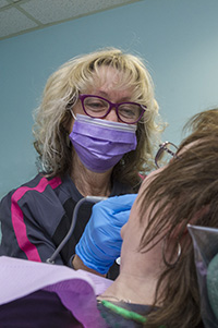
![]() Hygienist taking an intraoral photo of the patient’s mouth
Hygienist taking an intraoral photo of the patient’s mouth
- Intraoral camera—This pen-sized, camera-tipped wand, which we insert into your mouth, enables you to see the dental problems that require treatment. The intraoral camera takes photos of the inside of your mouth and transmits the images of your teeth through a computer unit to a large video screen. The images take some of the mystery out of dental treatment, enabling you to see the leakage around fillings and the fractures of teeth.
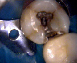
![]() Intraoral image showing decay underneath the old filling
Intraoral image showing decay underneath the old filling
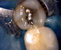
![]() Intraoral image showing a yellow area indicating early decay
Intraoral image showing a yellow area indicating early decay
The silver filling prevents the x-ray from showing any decay in its vicinity, and the early decay between the teeth is too narrow to be detected by x-ray.
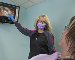
![]() Hygienist using a procedure video to explain treatment to the patient
Hygienist using a procedure video to explain treatment to the patient
- Patient education videos—We know that “a picture is worth a thousand words,” so we use procedure videos to show you your dental treatment options.
Office location
Dr. Fred S. Maron
541 Haight Avenue
Poughkeepsie, New York
12603
Phone: (845) 454-0380
FAX: (845) 454-2320
E-mail: contact@marondental.com
Wheelchair accessible
Our office has a wheelchair-accessible entrance and bathroom.
Testimonial
"I have been to other dentists over the past many years, but I think Dr. Maron is absolutely the best. No pain. Modern techniques. Superclean office. Wonderful staff."
Did you know?
The waterlines for our drills use a special additive to kill any bacteria in the waterline.
Testimonial
"He is constantly updating his equipment and techniques. I currently live in the City and come back to see him because he is the best."


